Proton Minibeam Radiation Therapy Shows Promise for Treating Glioblastoma
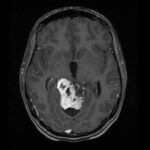
Radiation therapy (RT) is one of the most common and effective cancer treatments, used in over half of cancer patients. But for some tough cancers, like glioblastoma multiforme (GBM), RT has its limitations. This is because these tumors often have areas with low oxygen (hypoxia), suppress the immune system, resist radiation, and are close to…
COVID-19 Lung Damage Measurement
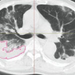
COVID-19, the infectious disease caused by the SARS-CoV-2 virus, continues to impact our daily lives. For those who become seriously ill with severe COVID-19 and require hospitalization, a severe pro-inflammatory condition can cause lung injury via pneumonia, acute respiratory distress syndrome, or sepsis. As the lungs and heart work together in the body to maintain…
Segmentation and Object Maps
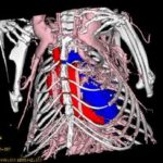
Segmentation is one of the most important functions of 3D image visualization and analysis. The goal of segmentation is to partition an image into regions, or objects, that are homogeneous with respect to one or more characteristics or features. Segmentation is a critically important tool for the processing of image data and is often the…
MicroCT Analysis of COPD-Related Bone Loss
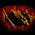
Preclinical imaging using microCT is a critical part of the drug discovery process, and allows significant insights into oncology, cardiovascular diseases, pulmonary diseases and orthopedics. While it is known that chronic obstructive pulmonary disease (COPD) and bone loss are related, almost all of the pathological mechanisms of COPD-related osteoporosis are unknown. A recent study by researchers…
Reducing CT radiation dose in liver cancer imaging
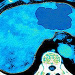
Recent improvements in the diagnosis and treatment of hepatocellular carcinoma (HCC), the most common type of primary liver cancer, have resulted from the use of CT perfusion imaging. This technique provides better tumor characterization by quantifying hepatic arterial blood flow as well as the morphology of the tumor. One of the drawbacks of this technique…
Adipose Tissue Classification and Quantification from MR and CT Imaging
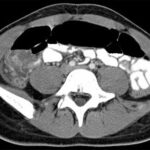
The study of adipose tissue is motivated by the negative consequences of obesity. Countless studies have reported that obesity is associated with an increased risk of heart disease, diabetes and other gastrointestinal diseases, and certain cancers. A search of the term “fat” in the Analyze Publications Database returns over 150 fat-related research articles in which…
3D Visualization of Subdural Electrodes
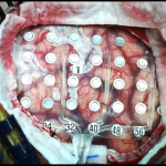
Intracranial EEG (iEEG), also known as electrocorticography or ECoG, is an invasive procedure used to place grid or strip electrodes on the surface of the brain to record electrical activity. From a clinical perspective, this procedure is extremely useful in evaluating patients with intractable epilepsy, that is, epilepsy that is unresponsive to anticonvulsants. These patients…
Image Registration and Fusion
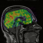
Registration is a 3D imaging process that allows information from different scanning modalities (for example MRI and PET) to be combined into one image space. The goal of image registration is to determine a spatial transformation that will bring into alignment separately acquired images of the same object. When accurately registered, each separate image will…
 AnalyzeDirect