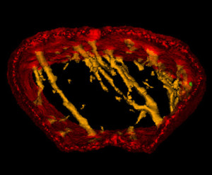
 Preclinical imaging using microCT is a critical part of the drug discovery process, and allows significant insights into oncology, cardiovascular diseases, pulmonary diseases and orthopedics.
While it is known that chronic obstructive pulmonary disease (COPD) and bone loss are related, almost all of the pathological mechanisms of COPD-related osteoporosis are unknown. A recent study by researchers in Japan established a COPD/emphysema-related osteoporosis mouse model by using elastase-induced emphysema.
A critical part of the study involved microCT scanning of the mouse model and determining key trabecular bone measurements. Analyze and the Bone Microarchitecture Analysis Add-On were used to establish bone microstructure parameters in the trabecular bone of L1 and the distal femoral metaphysis were evaluated. Specific parameters included trabecular bone volume (BV/TV: %), trabecular thickness (Tb.Th: mm), trabecular separation (Tb.Sp: mm), trabecular number (Tb.N: 1/mm), structure model index (SMI), and connectivity density (Conn.D: 1/mm3).
The study found that the lumbar vertebrae and femurs/tibiae of these models exhibited trabecular bone loss and impaired osteogenic activity in 24-week-old male elastase-induced emphysema model mice. In addition, the model mice showed atrophy of type I muscle fibers without atrophy of type II muscle fibers. The researchers concluded that emphysema may systemically induce bone/type I fiber loss and impair bone formation, and that the model they developed may be useful as a COPD/emphysema-related osteoporosis model in future drug discovery research.
Download Analyze 14.0 and the Bone Micro Analysis Add-On
 AnalyzeDirect