Bone Microarchitecture Analysis for MicroCT Data
Bone Microarchitecture Analysis Workflow
Preprocessing
The BMA add-on module requires that bone specimens are isovolumetric. If the image data contains multiple bones the bone of interest must be isolated and orientated into the correct anatomical orientation. All tools needed for data preprocessing are provided with Analyze.
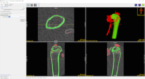
Cortical Bone Segmentation
Once the image data is loaded the BMA add-on will conduct a preliminary segmentation which will allow the module to identify the cortical shell, cortical pores and trabecular tissue regions.
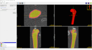
Trabecular Bone Segmentation
Upon user approval the add-on will now complete the segmentation process by identifying the trabecular bone in the trabecular region. Two new regions, the trabecular bone and intra-trabecular space will be derived.
Calculate Bone Measurements
Measurements are automatically calculated for the trabecular and cortical bone and output in several comma-separated value (.csv) files. A utility is also provided to convert CT numbers (HUs) to Bone Mineral Density (BMD) in units of mg/CC.
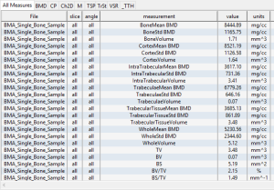
Table 1. Trabecular Bone Measurements
| Abbreviation | Description | Unit |
| TV | Total Volume | mm3 |
| BV | Bone Volume | mm3 |
| BS | Bone Surface | mm2 |
| BV/TV | Bone Volume Fraction | % |
| BS/TV | Bone Surface Density | mm2/mm3 |
| BS/BV | Specific Bone Surface | mm2/mm3 |
| Conn.D | Connectivity Density | 1/mm3 |
| SMI | Structure Model Index | |
| Tb.N | Trabecular Number | 1/mm |
| Tb.Th | Trabecular Thickness | mm |
| Tb.Sp | Trabecular Separation | mm |
| Tb.Th.SD | Standard Deviation of Trabecular Thickness | mm |
| Tb.Sp/SD | Standard Deviation of Trabecular Seperation | mm |
| DA | Degree of Anisotropy | |
| MIL | Mean Intercept Length |
Table 2. Cortical Bone Measurements
| Abbreviation | Description | Unit |
| Tt.Ar | Total Cross-Sectional Area | mm2 |
| Ct.Ar | Cortical Bone Area | mm2 |
| Ma.Ar | Medullary Area | mm2 |
| Ct.Ar/Tt.Ar | Cortical Area Fraction | % |
| Ct.Th | Average Cortical Thickness | mm |
| Ps.Pm | Periosteal perimeter | mm |
| Ec.Pm | Endocortical Perimeter | mm |
| Iap | Moment of Inertia Anteroposterior Axis | mm4 |
| Iml | Moment of Inertia Mediolateral Axis | mm4 |
| Imax | Maximum Moment of Inertia | mm4 |
| Imin | Minimum Moment of Inertia | mm4 |
| J | Polar Moment of Inertia | mm4 |
| Ct.Po | Cortical Porosity | % |
| Po.N | Pore Number | n |
| Po.V | Total Pore Volume | mm3 |
| AvgPo.V | Average Pore Volume | mm3 |
| Po.V.SD | Standard Deviation of Pore Volume | mm3 |
| Po.Dn | Pore Density | 1/mm3 |