 Recent improvements in the diagnosis and treatment of hepatocellular carcinoma (HCC), the most common type of primary liver cancer, have resulted from the use of CT perfusion imaging. This technique provides better tumor characterization by quantifying hepatic arterial blood flow as well as the morphology of the tumor. One of the drawbacks of this technique is the relatively high radiation dose resulting from repeated CT scanning of the liver.
Researchers in London, Ontario, investigated using much lower CT doses when scanning HCC patients by reconstructing undersampled CT liver images using compressed sensing or filtered backprojection techniques.
Five different sets of reconstructed CT images of the same patient were generated using various dose and reconstruction techniques, from the conventional full dose (984 views per 360 degrees) with filtered backprojection, to a third reduced dose (328 views) and quarter reduced dose (246 views), both datasets reconstructed with filtered backprojection, and both datasets reconstructed with compressed sensing.
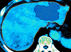
Using the Analyze software, each set of reconstructed DCE (dynamic contrast enhanced) liver images were aligned using the manual registration tools available in the Register module. Manual image registration is the process of aligning two or more images of the same space. The process involves designating one image as the base or reference image, and then using manual tools to applying geometric transformations to interactively translate and rotate the other match images so that they align with the base image. Once aligned, the match images are then transformed to the base image and saved. In this case, the saved registered images were exported into a perfusion analysis software program to determine what effects the scanning/reconstruction protocol had on the diagnostic quality of the liver anatomical images as well as the hepatic arterial blood flow measurements.
Comparing results for each of the five image sets concluded that the low CT dose (328 views) gives as good information as the conventional, higher dose protocol, suggesting that the dose to the patient can be reduced by up to 67% without impacting diagnostic quality. More HCC studies are needed to confirm the usefulness of this reduced dose protocol.
Tags: CT, Liver, Register, Tumor