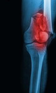
 Bones are made of living and growing tissue that is constantly being broken down and replaced. Osteoporosis is a disease that occurs when bone is removed too quickly or replaced too slowly, resulting in a gradual loss of bone mass, and a subsequent increased fragility that predisposes patients to elevated risk of fracture.
Fracture healing is a delicate and complex physiological process, requiring adequate stability and blood supply. Recent studies indicate that the reduced healing capacity of osteoporotic bones might be due to defects in mesenchymal stem cells that impair their ability to become osteoblasts, the major cellular component of bone.
However, differences in bone healing between osteoporotic and physiological bone remain uncertain. In a recent study, scientists from the University Hospital of Giessen-Marburg, Germany, investigated the influence of osteoporosis on bone healing patterns by inducing an osteoporotic bone status in 14 female rats.
Osteoporosis was caused by means of ovariectomy, the surgical removal of ovaries. This procedure causes a decrease in estrogen levels, a hormone produced by the ovaries that helps protect against bone loss. The rats were fed a diet low in calcium, which is essential to building strong, dense bones. A control group only underwent a faked surgical intervention that omits the step thought to be therapeutically necessary and therefore isolates the incidental effects caused by anesthesia, the incisional trauma and pre- and postoperative care. Three months later, an osteotomy – a surgical procedure whereby a bone is cut to shorten, lengthen, or change its alignment – of the left distal femur was performed in both groups.
After a six week healing period, femur specimens from each group were analyzed for discrepancies. Biomechanical testing revealed significant lower flexural rigidity in the osteoporosis group compared to the control group, a feature that reflects the local mechanical competence of the healing zone. Histological assessment of the fracture gap showed healing divergences such as cartilaginous remnant and more unmineralized tissue in the osteoporosis rats, compared to more mature consolidation appearance in the control group. Both results are in accordance and indicate delayed healing in the osteoporosis group. On the other hand, micro-CT analysis of new bone and vascular formation reflected no differences between the two groups, which may reflect the overall biomechanical stabilty met with the proposed fracture model. Analyze software was used to assess and measure cross-sectional areas of new ossified tissue formation and vascular volume fraction within the fracture gap.
These results support the clinical supposition of impaired healing in osteoporotic fractures. Future studies following a similar approach may be useful for the evaluation of potential agents for the prevention of post-menopausal osteoporosis.
Download our Guide to Bone Mineral Density Calibration
Tags: Bone Fracture, Orthopedic