 Orthopedic implant infections represent a severe complication following surgical procedures. This type of infection occurs by bacterial contamination either during surgery or immediately thereafter and leads to persistent inflammation, osteomyelitis (infection of the bone) and eventual osteolysis of the bone (autoimmune reaction which causes resorption of bone tissue). Very often these responses ultimately result in implant loosening and failure.
Studies have shown that whereas a minimum of about 10,000 bacteria are normally required to cause an infection in non-implant situations, as few as 10 can trigger an infection in the presence of an implant and once bacteria adhere to its surface, they become up to 1,000 times more resistant to antibiotics than systemic bacteria.
The diagnosis of infection is based on a combination of clinical signs, laboratory findings and imaging studies. Following this line of thinking, Bernthal et al. decided to monitor this biological phenomenon using a combination of imaging techniques. Their protocol consisted of in vivo bioluminescent and fluorescent imaging co-registered with micro-computed tomography imaging (micro-CT) of a mouse model of an orthopaedic prosthetic joint infection. The bioluminescence and fluorescence signals were produced by a bioluminescent strain of Staphylococcus aureus and the EGFP-fluorescent neutrophils of LysEGFP mice, respectively.
In order to investigate protective immune responses and pathologic anatomical changes, a state of infection was induced in the mice by pipetting an inoculum of bioluminescent Staphylococcus aureus onto the surface of the implant in the knee joint. In vivo optical imaging allowed the detection and quantification of the bioluminescent signal deriving from the bacteria, which belonged to a genetically engineered bacterial strain that constitutively emits light as a result of metabolic reactions. Meanwhile, in vivo fluorescent imaging was used to monitor inflammatory response by following the recruitment of the fluorescent neutrophils from the circulation towards the site of the infection. The co-registration of both optical signals with micro-CT images showed their 3D location in the anatomical context of the post-surgical knee joints.
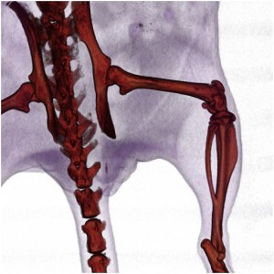
Analyze software was used to visualize and quantify the anatomical changes in quality and dimensions of the bone associated with the implant infection. 3D volumetric image analysis was performed using a semi-automated contour-based segmentation process and the results showed a substantial increase over time in the outer bone volume of the distal femur which was due to bone damage. For a detailed step-by-step guide of how to perform this procedure in Analyze, download the Volume Measurement and Rendering of Micro-CT Bone Data eBook.
Multimodality imaging allows a deeper understanding of the infection and inflammatory response and at the same time an evaluation of their consequences on the bone and joint tissue. This advance in noninvasive imaging of biological phenomena in the context of anatomical structures could potentially be expanded beyond infectious diseases and across disciplines to study other biological processes and pathological conditions.
Download our Guide to Volume Measurement
and Rendering of Micro-CT Bone Datat