 Chronically implantable devices take on an important role in the study of the functioning of the nervous system and have experienced a compelling evolution over the last decade. Cranial implants are used to electrically stimulate or record neural activity in the brains of awake behaving animal models.
The implantation of these devices often leads to complications that undermine the longevity and health of the implant itself and of the host animal. Common significant drawbacks encountered when using traditional cranial implants are infections, tissue damage, loosening of anchoring screws, skin recession, bone necrosis and diminished bone density given by bonding agents and extending support legs used to secure the device.
In order to minimize and prevent these shortcomings, scientists from McGovern Institute for Brain Research and Picower Center for Learning and Memory (MIT) developed innovative legless, custom-fit cranial implant devices.
Their improved implants are made from carbon-reinforced polyetheretherketone (PEEK), a biocompatible and radiolucent material which bestows optimal wear properties, load-bearing capacity and MRI-compatibility on the device. A key advance that has been developed to improve the implant design is to machine its base to be complementary in shape to the surface of the skull.
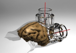
In order to create custom-fit cranial implants, the scientists derived the surface curvature of the cranium from MRI scans of non-human primates’ heads. Analyze software was used to segment the skull surface from the brain and skin and to design a 3D target vector that defined the desired position and orientation of the implant on the cranium. The customized implant designs were then used to machine the part from carbon-PEEK stock. The use of Analyze paved the way for a novel approach which allowed the researchers to perform the skull segmentation using just MR data, avoiding the need for acquisition and co-registration of CT data.
In these custom-fit cranial implants the screws and guide holes for the screws are encased within the walls of the chamber itself, reducing the overall device footprint and allowing for a smooth and uninterrupted interface between the edge of the device and the skin margin. This critical feature bypasses the need for bonding agents and extending support legs that commonly lead to tissue damage.
Moreover, these implants proved to be stable and suitable for reliable long-term recording and stimulation of neural activity, with no sign of infection. The technological advances that have been incorporated into the devices will lead to improved implant technology and be of benefit to the design of clinical applications for the treatment of neurological disorders.
Download our Guide to Skull Segmentation,
Vector Design and Surface Generation