 Temporal lobe epilepsy (TLE) is the most common form of partial or localized epilepsy in adults and accounts for approximately 60% of all patients with epilepsy. Medial temporal lobe epilepsy (mTLE), or limbic epilepsy, is the most common type of epilepsy, representing about 80% of all TLEs. mTLE is associated with head trauma, encephalitis, meningitis, birth injuries or malformations, and most commonly, a history of febrile seizures.
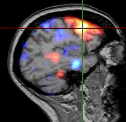
Difficult to treat, mTLE is often refractory to antiepilepsy medications. A common, but sporadic, finding on MRI is hippocampal sclerosis, with decreased hippocampal volume and cell loss. Additionally, decreased GABAergic inhibitory effect is thought to play a key role in the pathophysiology of the disease, but it is entirely clear whether this is related to cell loss, receptor loss, or changes in receptor properties. Decreased density of GABAA /central benzodiazepine receptors (GABAA/cBZR) in animals and humans with TLE is a well-established result seen on neuroimaging using radiolabeled receptor antagonist, flumazenil (FMZ) and PET.
FMZ/PET studies have conclusively shown decreases in GABAA/cBZR binding in the hippocampus on the same side as the epileptogenic focus. In studies looking at PET-detected receptor loss correlated with volumetric MRI, decreases in GABAA/cBZRs are greater than hippocampal volume loss, suggesting that the receptor loss is not completely explained by cell loss.
Traditional Model has Challenges for Routine Laboratory Use
Routine preclinical and clinical research using the standard radio-ligand, [C11]-FMZ, with PET has been hampered by the short half life of [C11]-FMZ and the need for multiple arterial sampling. However, recent modifications to the kinetics model and use of [18F]-FMZ, a radioligand with longer half life, are showing promising results for such applications.
Australian researchers [Vivash 2014] were interested in exploring the cell loss vs receptor change question by correlating receptor changes with neuronal degeneration using the new radio-ligand, [18F]-FMZ, and PET in rats. They adapted the [18F]-FMZ model to determine the concentration (Bmax) and binding affinity (1/KdVr) of GABAA /cBZRs in the standard post-kainic acid status epilepticus rat model.
Methods
First, researchers had to validate a modification of the PET model, using a partial saturation approach, in healthy rats. For this phase, PET scans were acquired using 3 injections of [18F]-FMZ with arterial sampling. PET images were analyzed using Analyze software and co-registered with an MRI template delineating areas of interest: whole brain, bilateral hippocampi, and olive nucleus.
After successful validation of the model, the modified [18F]-FMZ PET methodology was used to assess receptor density and affinity in kainic acid-induced status epilepticus in rats. Post- epilepsy-induction volumetric MRI was conducted, followed by [18F]-FMZ PET. Brain regions on MRI included hippocampus, amygdala, thalamus, olive nucleus and ventricles. PET series and MRI were co-registered and analyzed using Analyze software.
Results
The multi-injection partial saturation protocol [18F]-FMZ in rats successfully allowed for accurate kinetic modeling. Results of the study were conclusive for a significant decrease in GABAA /cBZR density in epileptic compared to control rats. However, a significant increase in hippocampal receptor binding affinity in the epileptic rats was observed. The decrease in receptors (Bmax) is consistent with similar results reported in humans with TLE. The increase in binding affinity (1/KdVr) was thought to be a disparate result of indirect measurement, 1/KdVr being a measure of the distribution of radiotracer rather than a direct measure of binding affinity.
While no overall differences were seen on MRI volumes between epileptic and control rats, hippocampal volume measures correlated with cell loss in the CA1, CA3 and CA3c areas in individual animals. Lack of correlation between receptor density and hippocampal volume or cell loss in rats is consistent with findings in human epilepsy patients, and confirms earlier observations that receptor changes are not entirely explained by cellular loss.
Conclusion
This is the first study using [18F]-FMZ PET to quantify GABAA /cBZR density and binding affinity in an in vivo animal model of epilepsy. Validation of the partial saturation model using [18F]-FMZ clears the way for more routine use of this model in longitudinal receptor studies without the necessity of multiple injections and serial arterial sampling. The authors were hopeful that these advancements would not only further illuminate the pathophysiology of TLE, but also facilitate the future use of [18F]-FMZ PET in clinical applications.
Download our Guide to Hippocampal Volume Assessment
Tags: Brain Studies, Epilepsy