Extra-Frontal Alterations in Juvenile Myoclonic Epilepsy
Several studies using state-of-the-art imaging techniques have revealed that juvenile myoclonic epilepsy, a form of epilepsy that starts in childhood or adolescence, might be associated with microstructural and functional changes in several brain regions. In fact, these abnormalities have been found not only the thalamofrontal network, but also in temporal and parieto-occipital areas. To further understand these…
Magnetic Resonance Spectroscopy for the Assessment of Temporal Lobe Epilepsy

Magnetic resonance imaging (MRI) offers valuable structural information about organs and tissues in the body, providing high-resolution images that help diagnose a wide range of conditions and dysfunctions. However, data underlying functional activity of the structure is often limited. Proton magnetic resonance spectroscopy (MRS) is a non-invasive, in vivo technique complementary to MRI that interrogates…
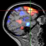
Differentiating Temporal Lobe Epilepsy through Functional Neuroimaging
Epilepsy is a brain disorder in which the normal pattern of neuronal activity becomes disturbed, causing changes in behavior, sensation or awareness and sometimes convulsions, muscle spasms and loss of consciousness. These symptoms, which go under the umbrella of seizures, may last from a few seconds to a few minutes. This disorder may result from events…
Temporal Lobe Epilepsy and Hippocampal Volume Loss – Cell Loss or Receptor Change?
Temporal lobe epilepsy (TLE) is the most common form of partial or localized epilepsy in adults and accounts for approximately 60% of all patients with epilepsy. Medial temporal lobe epilepsy (mTLE), or limbic epilepsy, is the most common type of epilepsy, representing about 80% of all TLEs. mTLE is associated with head trauma, encephalitis, meningitis,…