 Stroke is the second leading cause of death worldwide according to the World Health Organization. A stroke involves the loss of brain function due to a disturbance in blood supply to the brain. It can be caused by either a blood clot that blocks or plugs a blood vessel in the brain (ischemic stroke) or a blood vessel that breaks and bleeds into the brain (hemorrhagic stroke).
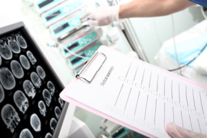
The NIH Stroke Scale (NIHSS) is a tool used to assess whether the degree of impairment caused by a stroke merits a specific type of treatment. This scale measures several aspects of brain function including vision, sensation, consciousness, movement and speech. A certain number of points are assigned for each impairment uncovered during a focused neurological examination and a maximum score of 42 represents the most severe and devastating stroke.
Advanced neuroimaging techniques also provide information that may be useful to guide treatment decisions. Diffusion-weighted imaging (DWI) has been shown to be highly sensitive for the diagnosis of cerebral infarction and for the identification of areas of acute infarcts, which appear hyperintense on diffusion images. In DWI scans, the intensity of each image element reflects the rate of water diffusion in that location, allowing the detection of areas in which water is unable to diffuse. Infarcts represent the core of irreversible damage and their analysis is thus crucial in selecting the right therapeutic guidelines to follow.
Mean transit time (MTT) maps are another sensitive and specific tool used for differentiating ischemic penumbra – the portion of the ischemic territory that is still potentially salvageable – from infarct core in acute stroke. MTT is a hemodynamic parameter that defines the time a certain volume of blood spends in the cerebral capillary circulation. MTT changes strongly correlate with the size of the ischemic penumbra, which is in turn typically operationally defined as the MTT lesion volume.
Scientists from Harvard Medical School recently published in the American Journal of Neuroradiology a predictive model that combines the NIHSS with MR imaging thresholds. DWI and MTT lesion volumes were matched with NIHSS score thresholds to predict outcome in patients who suffered from ischemic stroke.
Visually detected DWI and MTT abnormalities were segmented using Analyze software in order to obtain quantitative measurements of lesion volumes. Clinical and imaging data were compared and patients with DWI lesions of > 70 mL or an NIHSS score of > 20 had a high likelihood of poor outcome, while on the other hand MTT lesions of < 50 mL or an NIHSS score of < 8 predicted good outcome with a high positive predictive value.
Although further work is needed, these findings have potential implications for identifying the most beneficial and appropriate therapy for each patient.
Tags: Brain Lesions, Brain Studies, Stroke