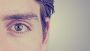
 Horner syndrome is a combination of signs and symptoms caused by the disruption of a nerve pathway that goes from the brain to the face and eye. The classical clinical findings associated with Horner syndrome are decreased pupil size (miosis), drooping eyelid (ptosis) and decreased sweating on the affected side of the face.
This condition is the result of other medical problems, such as stroke, tumor, spinal cord injury or carotid artery stent placement. There is no specific therapy for Horner syndrome, but treatment for the underlying cause may restore normal nerve function.
Scientists from Zeenat Qureshi Stroke Institute, St. Cloud, have recently published a case study describing Horner syndrome in a patient after left carotid artery stent placement. This endovascular procedure consists of the implant of a wire mesh stainless steel tube that becomes a permanent part of the artery, holds it open and keeps it from closing again. After surgery, the subject developed miosis and ptosis of the left eye, symptoms strictly associated with Horner syndrome. The investigators performed a combination of imaging techniques and pharmacological testing in order to localize the nerve lesion causing the condition. In fact, disruption of the nerve pathway may involve either pre-ganglionic neurons or post-ganglionic neurons. The pre-ganglionic ones arise in the central nervous system and run to a ganglion in the body. Here they synapse with postganglionic neurons, which in turn run to the effector organ (cardiac muscle, smooth muscle, or a gland).
The group was able to distinguish between pre- and post-ganglionic lesion measuring pupillary responses to drops of phenylephrine in the conjuctival sacs. Phenylephrine is a mimetic drug – a compound used to mimic the effects of norepinephrine, the neurotransmitter released by the post-ganglionic neurons that supply the iris dilator muscle and cause increase pupil size.
It has been known for many years that denervation of an organ leads to an increase in its responsiveness to the corresponding neurotransmitter or to a chemically related compound – a concept that goes under the name of “denervation supersensitivity”. Thus, a lesion interrupting the post-ganglionic fibers – causing therefore denervation of the iris dilator muscle – should considerably dilate the pupil when phenylephrine is placed in the eye. On the other hand, pupils may only dilate minimally with pre-ganglionic lesions.
The researchers photographed both pupils at baseline and post-treatment. The images were exported into Analyze software and their diameter was measured. The affected eye demonstrated a minimal increase which allowed the group to categorize the lesion as a pre-ganglionic one. This study offers pharmacological testing for the identification of post carotid stent Horner syndrome and provides additional information that may help characterize the underlying pathological cause of this condition.
Tags: vision