 Imagine how difficult it would be to lose your voice, to be unable to convey your thoughts and needs via speech. It may be more likely than you think. Approximately 6 percent of the US population develops a voice disorder during their lifetime. Singers, teachers, and politicians are obvious examples of occupations prone to overused vocalization that results in vocal fold abnormalities. But more inclusively, we are all at risk, given our extended life expectancy. The older we get, the longer we use our voice, the more likely it is for some of us to experience vocal fold damage, commonly scarring, leading to weakened and raspy speech. Vocal chord pathologies: nodules polyps, ulcers, laryngitis, cancers, paralysis and paresis all count on research to find preventions and treatments for vocal health.
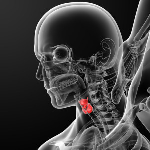
Vocal Fold Structure and Mechanics
The vocal folds modulate air flow through the larynx during vocalization. They are composed of an epithelial mucosa supported by an extracellular matrix (ECM) of fibrous and interstitial proteins and glycosaminoglycans. The density and arrangement of the ECM molecules is very important for the folds to retain both tensile strength and elasticity such that they can vibrate as breath flows through them. Changes in the shape, size, or constituents of the ECM can have dramatic effects on vocalization.
Problems with Quantifying Abnormal Vocal Fold Changes
Research on vocal folds involves intricate knowledge of the larynx and its associated structures. Ex vivo processing of larynges for characterization of the vocal folds is troublesome, as all available methods, freezing, resection, formalin fixation and staining impose some artifact on vocal fold geometry. However, if a very specific determination of the degree of artifact can be measured, then this can be taken into account, allowing for accurate and consistent geometric evaluation.
Improved Measurement of Laryngeal Geometry
Efforts in acoustical research have been aimed at improving definition of laryngeal anatomical geometry, as well as accurate measurement of processing artifact. Advanced bio-imaging technologies are playing a major role in model development for the characterization of vocal mechanics.
For example, Tayama et al in 2000 photographed and measured consecutive coronal vocal fold sections, taken after quick-freezing the tissue, and measured after thawing in saline. Geometric change as a result of freezing was a 5% expansion, followed by a 1-2% reduction to pre-frozen size on thawing. They concluded that the thawed measurements were good approximations of in vivo vocal fold dimensions.
A more recent study presented at the 2013 International Congress on Acoustics (ICA) by Stevens et al builds on Tayama’s findings, but using 3-dimensional imaging and volume determination to further characterize laryngeal fresh-freeze volume discrepancies. As opposed to canine vocal folds, which have traditionally been employed in phonetics research, these scientists employed porcine larynges, which are anatomically similar to human and are readily available.
Comparison of pre- and post-frozen laryngeal volume was made from microCT scans. The fresh laryngeal tissue was scanned, then frozen with liquid nitrogen-cooled isopentane and rescanned. Analyze was used to create a volumetric representation of each larynx from both fresh and frozen tissue images.
All larynges tested had a post-freeze volume expansion of greater than 3 percent, with approximately a 3.5% mean. In addition, the airway volume was consistently larger in frozen larynges. The researchers hoped that these results would be useful as a baseline for ongoing laryngeal geometric volume measurements.
The future study of phonetic research will undoubtedly find great benefit through correlation of 3-dimensional geometric vocal fold measurement with histological tissue changes.
Tags: Larynx, Vocal Fold Geometry