 A precise view inside coronary arteries speaks volumes about the health of a patient’s heart. This is particularly true when it comes to detecting the extent of life-threatening plaques inside coronary arteries and dangerous left arterial bifurcation angles. A recent study has found that coronary CT virtual intravascular endoscopy can be used to identify different plaque types, while providing detailed 3D views of the intraluminal surfaces and geometry of the coronary arteries.
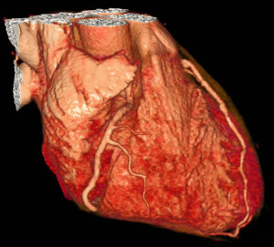
Healthy vs. unhealthy arteries
It has been common knowledge for decades that cholesterol plaques clog coronary arteries inside the heart, potentially leading to heart attack. It may be less commonly known that the tilt of heart’s left arteries, referred to as left arterial bifurcation angle, negatively affects the flow of blood through coronary arteries, which stresses the heart.
A look inside a healthy heart would show a smooth intraluminal appearance of the coronary artery walls. A heart with coronary artery disease would show irregular-shaped plaque bulges protruding from the coronary artery walls. A look inside the heart would also reveal the tilt of left arterial bifurcation angles. The marker for the presence of coronary artery disease is left arterial bifurcation angles greater than 80 degrees.
What 3D virtual images reveal
At Curtin University in Perth, Australia, medical imaging specialist Zhonghua Sun recently applied 3D CT virtual intravascular endoscopy to visualize exactly what was happening inside the coronary arteries of 50 patients suspected of having coronary artery disease. The patients included 31 men and 19 women who were approximately 56 years old. The findings of his research appeared in a recent edition of BioMed Research International.
For the study, CT scans were collected on these 50 patients who had recently undergone CT angiography. The purpose of this retrospective study was to apply 3D CT virtual intravascular endoscopy diagnostically to view the intraluminal appearances of coronary plaques, examine plaque components and calculate left coronary bifurcation angles. Another key purpose was to use 3D CT virtual intravascular endoscopy to learn whether there is a connection among coronary wall appearances, left bifurcation angles and different types of plaques.
To create detailed visualization of coronary arteries, the patients’ coronary CT angiographs were reconstructed into 3D virtual intravascular endoscopy images using Analyze. The end product was a view of the intraluminal appearance of coronary artery walls, the variety of plaques present in coronary arteries and left coronary bifurcation angles. These 3D virtual images also enabled comparisons of plaque components and left coronary bifurcation angles in coronary arteries.
What coronary artery disease looks like
The 3D virtual images revealed that the greater the left arterial bifurcation angle, the greater the coronary artery disease. Also, distinct irregular-shaped plaque bulges in coronary artery walls indicated the extent of coronary artery disease.
In this specific study, the left arterial bifurcation angles in 25 patients with diseased left coronary arteries tilted at 94.3 degrees. This compares to 76.5-degree angles observed on 3D virtual images of the patients with normal left coronary arteries.
As for deadly plaques, the 3D virtual images showed the protruding, irregular regions on the coronary artery wall filled with plaques. These irregular regions were evident in 10 out of 11 patients with mixed plaques in left anterior descending arteries and left circumflex arteries. In 29 patients, the more dangerous calcified plaques were visualized in these same arteries. Bulging plaques in coronary artery walls were also found in approximately 50 percent of patients with unhealthy left bifurcation angles tilting more than 80 degrees.
A positive study finding was how healthy coronary arteries appeared. In 95 percent of the patients with suitable left bifurcation angles of less than 80 percent, the 3D virtual images showed smooth-appearing coronary artery walls with no bulging plaques.
Overall, the bottom line is that viewing the interior of coronary walls with 3D CT virtual intravascular endoscopy technology reveals with precision the condition of a patient’s heart. This investigation also demonstrated the promising use for 3D CT virtual intravascular endoscopy to assess coronary plaques, left bifurcation angles and other mechanisms of action in the coronary artery wall.
Tags: Coronary Artery Disease, CT Angiography, Virtual Endoscopy