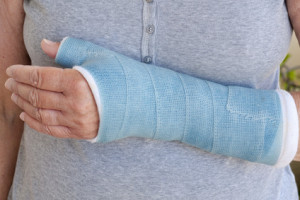
 Bennett’s fracture is the most common type of fracture of the thumb. It is a fracture of the base of the thumb which extends into the joint of the small bones of the hand. The fracture process involves the bone breaking away from the joint, leaving behind a small volar bone fragment, known as the palmar beak fragment, that remains attached to one of the ligaments, while the larger fragment subluxes or dislocates dorsally. This injury may be caused by striking something with the thumb extended, common with activities like martial arts, football, soccer or rugby.
Imaging tools such as Computed Tomography (CT) scans of carpometacarpal joints are usually performed to diagnose Bennett’s fracture. A specific type of CT called Computed Tomography – Osteoabsorptiometry (OAM) examines the distribution of subchondral bone mineralization which represents the load pattern, or pressure distribution, in the joint surface.
Scientists from the Department of Trauma Surgery and Sports Medicine in Innsbruck, Austria, decided to further investigate one of the methods of treatment for Bennett’s fracture: closed reduction followed by percutaneous K-wire fixation, which entails a post-operative step-off – an incongruence in the alignment of the bone fragments. The researchers were determined to understand whether this residual joint step and altered load distribution led to negative clinical outcomes or symptomatic degenerative osteoarthritis.
The 24 patients available for long-term follow-up examination who were included in this study had all previously undergone surgery and all presented a residual step-off in the articular surface at the region of the palmar beak fragment. CT scans of both carpometacarpal joints were performed and Analyze software was used as the image analysis system to conduct CT-OAM. In order to determine the areas of maximum density, the joint surface was virtually subdivided into 9 anatomic regions. The degree of mineralization within the joint surface was then measured for each region.
The highest degree of mineralization was found in the radial area of the carpometacarpal joint of the thumb – the joint that connects the trapezium to the first metacarpal bone – and no higher loading in the area of the palmar beak fragment (volar-ulnar area) could be found. This result suggested that a post-operative step-off may provide an overriding beneficial effect by leading to an unloading of the volar region, which is the primary contact area of the metacarpal beak.
The researchers found that this minimally invasive method is sufficient to reduce subluxation (incomplete or partial dislocation of the joint), provides the beneficial effect of pressure distribution while maintaining ligament support, and prevents the development of any major osteoarthritis in long-term investigations.
Tags: Bone Fracture, Osteoarthritis AnalyzeDirect