 Women with a shortened cervix during pregnancy are at high risk for premature delivery; the shorter the cervix, the higher the risk. An estimated 500,000 births, 12% of all births annually in the US, are preterm and end in significant infant mortality and morbidity. Associated healthcare costs are a whopping $26 billion per year.
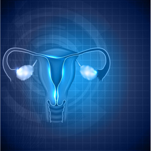
Cervical change is a natural progression of the body’s preparation for fetal passage. As parturition nears, the cervical tissues soften and hydrate, allowing for effacement and delivery. Monitoring cervical change is a standard part of obstetrical care and now, transvaginal ultrasound allows for accurate cervical measurement with pursuant interventions, if necessary. Treatment for prevention of preterm delivery when cervical shortening exceeds certain measures is accomplished with supplemental progesterone, cervical circlage or cervical pessary.
As common and costly as is preterm birth, it is reasonable to investigate related factors and mechanisms that facilitate such an event. Researchers in obstetrics and gynecology, biomedical engineering, and health science technology at MIT, Tufts University, and Tufts Medical Center are collaborating to more fully define the three-dimensional anatomical changes that lead to cervical shortening during pregnancy.
In their recently published study, MRI and 3-D ultrasound were used to accurately measure both uteran and cervical volumes during gestation. These technologies produced high resolution images which were converted for processing to a multidimensional imaging format using the Analyze software. T2-weighted and proton density MRI pulse sequences were registered and fused automatically with Analyze, whle the transperineal and transabdominal ultrasound images were manually registered and fused. The imaging data was then used to construct solid 3D models for anatomical visualization and further conversion to numerical models suitable for biomechanical analysis.
Cervical deformation in relationship to uteran change was examined. Cervical changes were most evident with inferior uteran growth and fundal pressure, which displaces both internal and external cervical os and initiates funneling. Changes at the internal os are particularly associated with coincident tissue changes in the cervical stroma. Interestingly, single fetus pregnancies displaced the interior cervical os anteriorly, while with twins, the interior os was displaced inferiorly. Cervical shortening was only observed with inferior displacement and in a ratio of 1cm shortening for 2cm of displacement.
The sequence of anatomical changes and histological counterpart during cervical funneling has been thoroughly described. Briefly, the extent of fetal membrane adhesion and continued uteran pressure promote cervical readiness for parturition. By simulating uteran growth as a result of gravitational loads, amnionic pressure, and uteran stretch, researchers were able to observe the related mechanisms involved in, and resulting from, lower uteran change. These included increasing gravitation load, stretching uteran walls, and changing geometry of uteran supporting structures: the endopelvic fascia, utero-sacral ligaments, cardinal ligaments and pelvic floor. Good correlation was found between the ultrasound-based cervical response and the numerical simulation.
Accurate timing of cervical function vis a vis fetal growth and membrane coherence is necessary for healthy pregnancy and full term delivery. While many factors can be involved in short cervix leading to preterm birth, the successful generation of numerical models from 2 and 3-dimensional in vivo images allows more discrete characterization of the synchronous biomechanical relationship between uterus and cervix in preparation for successful parturition. This information can improve treatment for ensuring cervical function, and potentially delaying preterm birth.
Tags: Cervical Shortening, Cervix, Pregnancy, Prematurity