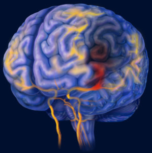
 Aging is associated with the appearance of increased white spots visualized on brain MRI scans. The presence of these anomalies, called white matter hyperintensities, characterizes elderly subjects as well as ischemic stroke patients, regardless of their age. White matter hyperintensities are thought to be caused by small vessel infarcts (restriction in blood flow) in the white matter and ultimately result in impairment of brain functions, such as cognition, balance and gait, that depend on complicated interactions between regions.
A recent study carried out by scientists from the Catholic University of Korea indicates that white matter hyperintensities in ischemic stroke patients may be subject to dynamic changes over time. The participants underwent brain MRI scans from which Analyze software was used to visually assess the hyperintensities and measure their volume. The differences in lesion volumes between the initial scans and the follow-up ones were interpreted as progression (increased volume), regression (decreased volume) or progression in particular areas of the brain and regression in others.
While about 32% of the patients developed new white matter hyperintensities over time, about 21% of them showed smaller lesion volumes. The reversibility of these abnormalities, that is, the observation that hyperintensities may not only progress but also regress, had never been observed in stroke patients in any previous study.
Spurred on by the novel findings of this study, researchers decided to search for clinical factors associated with each changing pattern. They found that individuals who showed increased white spots were older males with large vessel disease and decreased renal function. These two conditions are responsible for diffuse ischemia and small-vessel pathology, respectively, both risk factors for the development of white matter lesions.
Although no significant clinical variable was found to be associated with regression of lesion volumes, the results from this study indicate for the first time that there is a reversible mechanism responsible for the development of white matter hyperintensities over time. Further studies that investigate the variables that rule these dynamics are warranted.
Download our Guide to Intracerebral Hematoma Volume Measurement from CT
Tags: Aging, Brain Lesions, Brain Studies, Stroke